

 Tiếng Việt
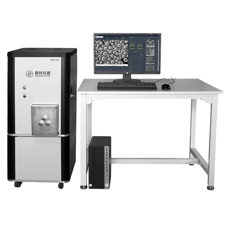
SS-150 Desktop Scanning Electron Microscope
Discover how a desktop scanning electron microscope (Desktop SEM) provides compact, high-resolution microstructure and surface analysis for modern laboratories.
Product Information
Desktop Scanning Electron Microscope: The Ultimate Guide to Compact Microstructure Analysis
Are you looking for a high-magnification surface analysis tool but facing limited laboratory space? A desktop scanning electron microscope (also known as a Benchtop SEM) is the perfect answer. This innovative instrument packs the immense analytical power of a traditional, floor-standing SEM into a compact, user-friendly, and cost-effective design.
In this comprehensive guide, we will explore the technology, core operational modes, key benefits, and diverse applications of this game-changing laboratory equipment.
1. What is a Desktop Scanning Electron Microscope?
A desktop scanning electron microscope is a miniaturized version of industrial electron microscopes. Instead of using visible light like a conventional optical microscope, a desktop SEM utilizes a highly focused beam of electrons to scan the surface of a sample.
By interacting with the atoms in the sample, the electron beam produces various signals that contain detailed information about the specimen’s surface topography and composition. Thanks to its streamlined engineering, the entire system can sit directly on a standard laboratory workbench, completely eliminating the need for specialized facilities or heavy-duty infrastructure.
2. Core Imaging Modes of Benchtop SEMs
Modern desktop scanning electron microscope systems are highly versatile, typically incorporating two primary electron detection modes to deliver comprehensive structural data:
Secondary Electron (SE) Mode
- The Mechanism: The SE detector captures low-energy electrons knocked out from the outermost, shallow layers of the sample surface by the primary electron beam.
- The Application: This mode is dedicated to topographical analysis. It generates high-resolution, three-dimensional-like images that reveal surface roughness, grain structures, fractures, pores, and the precise shapes of nanomaterials.
Backscattered Electron (BSE) Mode
- The Mechanism: The BSE detector records high-energy electrons that penetrate deeper into the specimen and bounce back. The intensity of this signal is directly proportional to the atomic number (Z) of the elements in the scanned area.
- The Application: This mode specializes in compositional contrast analysis. Regions composed of heavier elements (higher Z) appear brighter, while areas with lighter elements appear darker. This allows researchers to quickly differentiate between different material phases without destructive testing.
3. Why Modern Laboratories Prefer Desktop SEMs
The global demand for the desktop scanning electron microscope has surged rapidly due to several distinct operational advantages over traditional floor-standing units:
- Space-Saving Design: Its minimal footprint frees up valuable laboratory space. It does not require dedicated rooms with complex anti-vibration flooring or external water-cooling systems.
- Rapid Vacuum Cycle: Traditional SEMs take a long time to pump down. A desktop SEM achieves the required high-vacuum state in just 1 to 3 minutes, dramatically accelerating sample throughput.
- Intuitive User Interface: The software is designed with heavy automation. Features like auto-focus, auto-alignment, and auto-contrast allow novice operators to capture high-quality images with minimal training.
- Cost Efficiency: It offers a lower initial capital investment and significantly cheaper long-term maintenance, making electron microscopy accessible to small startups and academic labs.
- Seamless Upgradability: Most desktop systems can be integrated with an Energy-Dispersive X-ray Spectrometer (EDS/EDX). This allows for simultaneous qualitative and quantitative elemental mapping directly on the visual interface.
4. Practical Applications Across Industries
The versatility of the desktop scanning electron microscope makes it an indispensable tool across multiple sectors:
- Materials Science & Metallurgy: Examining metal fracture surfaces, analyzing thin-film coatings, identifying micro-defects, and inspecting advanced ceramics.
- Semiconductor & Electronics: Inspecting microchip defects, checking solder joints, and measuring the critical dimensions of nanoscale electronic components.
- Pharmaceuticals & Life Sciences: Observing the morphology of drug carrier particles, evaluating pill coatings, and investigating biological fibers or scaffolds.
- Energy & Battery Research: Analyzing the microstructure of electrode materials and investigating degradation mechanisms in Lithium-ion batteries.
5. Conclusion
The desktop scanning electron microscope bridges the gap between basic optical inspection and industrial-scale electron microscopy. It empowers laboratories to conduct rapid, high-resolution microstructure imaging and elemental analysis without the high costs and spatial constraints of traditional systems.
Be the first to review “SS-150 Desktop Scanning Electron Microscope”
Related products
ANALYSIS INSTRUMENT
ANALYSIS INSTRUMENT
ANALYSIS INSTRUMENT

ANALYSIS INSTRUMENT
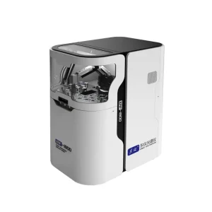
DF-1000E EDXRF Non-Destructive Elemental Composition Analyzer
ANALYSIS INSTRUMENT
ANALYSIS INSTRUMENT
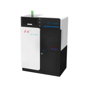
DF-1600 EDXRF The Construction Material Composition Analyzer
ANALYSIS INSTRUMENT

ANALYSIS INSTRUMENT







Reviews
There are no reviews yet.